Diagram Of Hip.and Back.muscles : Learn all muscles with quizzes and labeled diagrams | Kenhub / The extrinsic muscles that are associated with upper extremity and shoulder movement, and injuries of the intrinsic back muscles often occur while using improper lifting technique.
Diagram Of Hip.and Back.muscles : Learn all muscles with quizzes and labeled diagrams | Kenhub / The extrinsic muscles that are associated with upper extremity and shoulder movement, and injuries of the intrinsic back muscles often occur while using improper lifting technique.. The main muscles of the hip and pelvis consistsof the iliopsoas, pectinues, rectus femoris and sartorius at the front. Luckily you've found this page to help you. Other muscles are small and cover much less space. The levator ani muscle along with a second muscle forms the pelvic floor. Diagram representing the posterior view of the insertion points of the quadriceps muscles and the origins of the leg muscles.
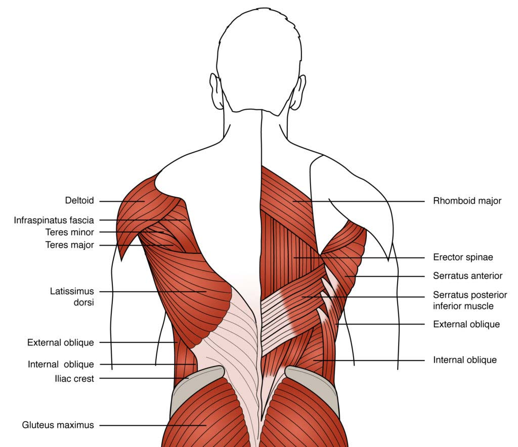
The back's muscles start at the top of the back (named the cervical vertebrae) and go to the tailbone (also named the coccyx). Back muscles anatomy lower back muscles anatomy human anatomy. It joins the lower limb to the pelvic girdle. It is opposite from the chest, and the vertebral column runs down. Human muscle system, the muscles of the human body that work the skeletal system, that are under voluntary control, and that are concerned with movement, posture, and balance.
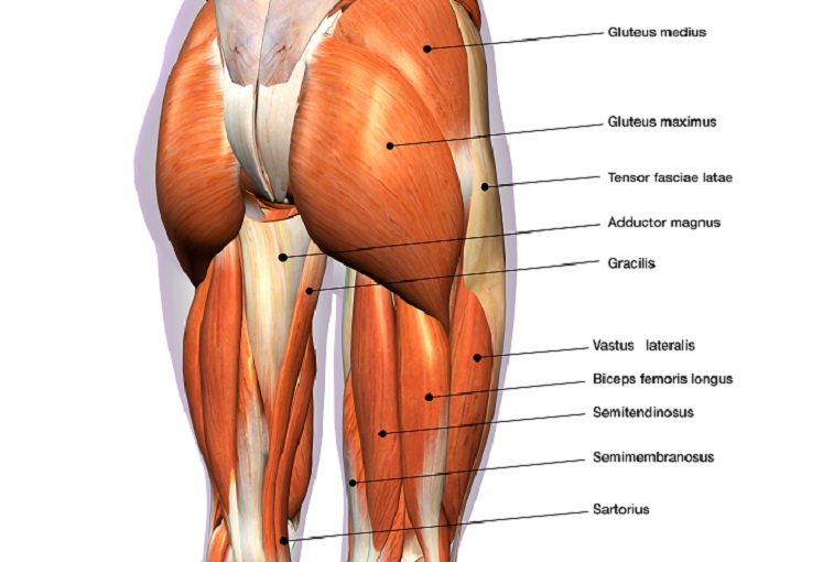
Pin on Medicina from i.pinimg.com All about the back muscles shares the back anatomy includes the latissimus dorsi trapezius erector spinae rhomboid and the teres lower back muscles diagram human back muscles anatomy on human. Body muscle structure 12 photos of the body muscle structure body muscle chart exercises, body muscle chart for bodybuilding, body muscle names chart, body muscle ratio chart, human body muscle chart free, human muscles, body muscle chart exercises. There are around 650 skeletal muscles within the typical human body. Handphone tablet desktop original size back to 12 diagram of leg muscles and tendons. Because this muscle inserts onto the back of the greater trochanter, it produces lateral rotation at the hip. The levator ani muscle along with a second muscle forms the pelvic floor. Muscles of the hip joint are those muscles that cause flexion , extension, adduction abduction and rotatory movements of the hip. The hip muscle diagram below shows a number of the muscles we will be discussing in the next sections.
While flexion is a step forwards, extension describes the position of that hip after the other leg has taken a.
In human anatomy, the muscles of the hip joint are those muscles that cause movement in the hip. Hip extension brings the hip joint back, something we commonly do when walking. Now that you watched the video, you. Muscles of hip bone and spine. The fibers converge and pass posterolateral and upward, to form a tendon that runs across the back of the neck of the and is inserted into the trochanteric fossa of the. Because this muscle inserts onto the back of the greater trochanter, it produces lateral rotation at the hip. Most modern anatomists define 17 of these muscles, although some additional muscles may sometimes be considered. The gluteus medius, gluteus minimus, piriformis, tensor fasciae latae on the outside. Related posts of muscles of the lower back and hip diagram muscle anatomy posterior. Abduction and medial rotation at the hip. The main muscles of the hip and pelvis consistsof the iliopsoas, pectinues, rectus femoris and sartorius at the front. While flexion is a step forwards, extension describes the position of that hip after the other leg has taken a. You can protect the back muscles by bending from the hip and.
Extension and lateral rotation at the hip. Now that you watched the video, you. This is a table of skeletal muscles of the human anatomy. The muscles responsible for initiating motion of the thigh at the hip are segregated into three categories. Muscles of hip bone and spine.
Why are core muscles important for back pain? | London ... from backpaindoctor.co.uk Muscles of hip bone and spine. The back's muscles start at the top of the back (named the cervical vertebrae) and go to the tailbone (also named the coccyx). All about the back muscles shares the back anatomy includes the latissimus dorsi trapezius erector spinae rhomboid and the teres lower back muscles diagram human back muscles anatomy on human. Some of these muscles are quite large and cover broad areas. Gluteus maximus, biceps femoris, semitendinosus, semimembranosus at the back and the. There are around 650 skeletal muscles within the typical human body. It joins the lower limb to the pelvic girdle. The levator ani muscle along with a second muscle forms the pelvic floor.
You can protect the back muscles by bending from the hip and.
• the sciatic nerve passes just inferior to the piriformis therefore a tight piriformis muscle my contribute to compression on the sciatic nerve. It is opposite from the chest, and the vertebral column runs down. The former two groups, superficial and intermediate, are referred to as the extrinsic back muscles. Now that you watched the video, you. The core muscles are those in the abdomen, back, and pelvis, and they also stabilize the body and assist in tasks, such as lifting weights. Extension and lateral rotation at the hip. The gluteus medius, gluteus minimus, piriformis, tensor fasciae latae on the outside. Diagram of muscles and anatomy charts. Related posts of muscles of the lower back and hip diagram muscle anatomy posterior. Dislocation of the hip joint. Want to learn more about it? All of these things can lead to long term back pain (and chronic complaining!). Gluteus maximus, biceps femoris, semitendinosus, semimembranosus at the back and the.
Gluteus maximus, biceps femoris, semitendinosus, semimembranosus at the back and the. Back muscles anatomy lower back muscles anatomy human anatomy. Extension and lateral rotation at the hip. It is opposite from the chest, and the vertebral column runs down. They are the biceps femoris (long head and short head), semimembranosus, and semitendinosus.
Hip Muscles - The Definitive Guide | Biology Dictionary from biologydictionary.net The achilles tendon attaches the muscles of the. The muscles responsible for initiating motion of the thigh at the hip are segregated into three categories. Body muscle structure 12 photos of the body muscle structure body muscle chart exercises, body muscle chart for bodybuilding, body muscle names chart, body muscle ratio chart, human body muscle chart free, human muscles, body muscle chart exercises. The former two groups, superficial and intermediate, are referred to as the extrinsic back muscles. Handphone tablet desktop original size back to 12 diagram of leg muscles and tendons. The main muscles of the hip and pelvis consistsof the iliopsoas, pectinues, rectus femoris and sartorius at the front. Extension and lateral rotation at the hip. Human muscle system, the muscles of the human body that work the skeletal system, that are under voluntary control, and that are concerned with movement, posture, and balance.
Want to learn more about it?
Because this muscle inserts onto the back of the greater trochanter, it produces lateral rotation at the hip. Related posts of muscles of the lower back and hip diagram muscle anatomy posterior. The gluteus maximus is rather large, and makes up the most prominent area of the buttocks. Learn with flashcards, games and more — for free. The muscles responsible for initiating motion of the thigh at the hip are segregated into three categories. Muscles of back of hip an… category: In human anatomy, the muscles of the hip joint are those muscles that cause movement in the hip. Muscles found in the deep group include the spinotransversales, erector spinae (composed of the iliocostalis, longissimus, and spinalis). The levator ani muscle along with a second muscle forms the pelvic floor. Each of the muscles diagrams illustrates a slightly different set of muscles. Most modern anatomists define 17 of these muscles, although some additional muscles may sometimes be considered. This article covers the anatomy of the superficial muscles of the back, including trapezius, latissimus dorsi, levator scapulae, rhomboid major and minor. Francesca salvador msc last + show all.


0 Komentar